品质至上,客户至上,您的满意就是我们的目标
技术文章
当前位置: 首页 > 技术文章
利用计算机断层扫描系统进行种子根系表型以及种质资源研究
发表时间:2021-08-16 14:38:00点击:1148
来源:北京博普特科技有限公司
分享:
近年来,科学家利用台式和落地式计算机断层扫描系统发表了多篇植物根系、种子表型无损研究研究的文章,文章发表在Frontiers in Plant Science,Plant Phenomics,Plant Methods,Journal of Imaging等主流期刊上。
北京博普特科技有限公司代理系列计算断层扫描系统,专注于该系统在植物、种子表型、土壤等领域的应用。


Direct comparison of MRI and X-ray CT technologies for 3D imaging of root systems in soil: Potential and challenges for root trait quantification
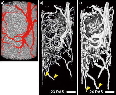
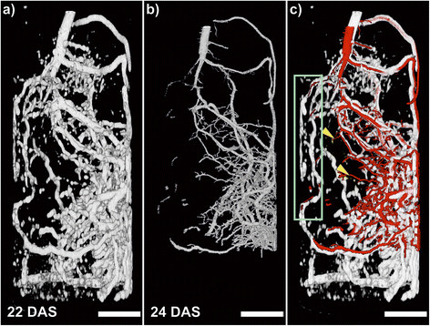
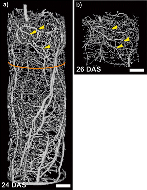
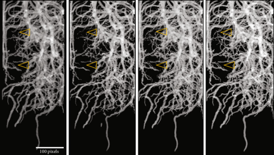
Roots are vital to plants for soil exploration and uptake of water and nutrients. Root performance is critical for growth and yield of plants, in particular when resources are limited. Since roots develop in strong interaction with the soil matrix, tools are required that can visualize and quantify root growth in opaque soil at best in 3D. Two modalities that are suited for such investigations are X-ray Computed Tomography (CT) and Magnetic Resonance Imaging (MRI). Due to the different physical principles they are based on, these modalities have their specific potentials and challenges for root phenotyping. We compared the two methods by imaging the same root systems grown in 3 different pot sizes with inner diameters of 34 mm, 56 mm or 81 mm. Both methods successfully visualized roots of two weeks old bean plants in all three pot sizes. Similar root images and almost the same root length were obtained for roots grown in the small pot, while more root details showed up in the CT images compared to MRI. For the medium sized pot, MRI showed more roots and higher root lengths whereas at some spots thin roots were only found by CT and the high water content apparently affected CT more than MRI. For the large pot, MRI detected much more roots including some laterals than CT. Both techniques performed equally well for pots with small diameters which are best suited to monitor root development of seedlings. To investigate specific root details or finely graduated root diameters of thin roots, CT was advantageous as it provided the higher spatial resolution. For larger pot diameters, MRI delivered higher fractions of the root systems than CT, most likely because of the strong root-to-soil contrast achievable by MRI. Since complementary information can be gathered with CT and MRI, a combination of the two modalities could open a whole range of additional possibilities like analysis of root system traits in different soil structures or under varying soil moisture.
Transfer Learning from Synthetic Data Applied to Soil–Root Segmentation in X-Ray Tomography Images
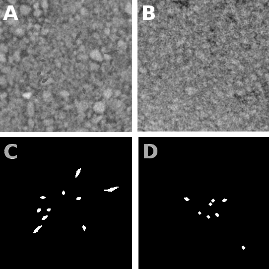


One of the most challenging computer vision problems in the plant sciences is the segmentation of roots and soil in X-ray tomography. So far, this has been addressed using classical image analysis methods. In this paper, we address this soil–root segmentation problem in X-ray tomography using a variant of supervised deep learning-based classification called transfer learning where the learning stage is based on simulated data. The robustness of this technique, tested for the first time with this plant science problem, is established using soil–roots with very low contrast in X-ray tomography. We also demonstrate the possibility of efficiently segmenting the root from the soil while learning using purely synthetic soil and roots. © 2018 by the authors. Licensee MDPI, Basel, Switzerland. This article is an open access article distributed under the terms and conditions of the Creative Commons Attribution (CC BY) license
Quantification of seed performance: non-invasive determination of internal traits using computed tomography
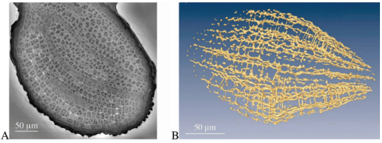
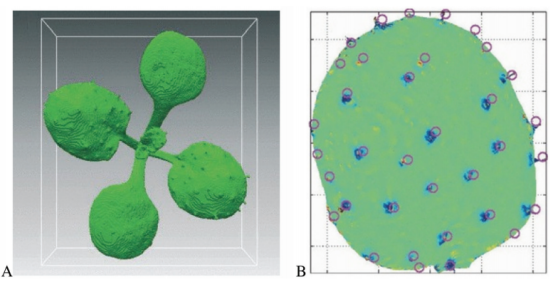
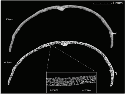
The application of the 3D mean-shift filter to 3D Computed Tomography Data enables the segmentation of internal traits. Specifically in maize seeds this approach gives the opportunity to separate the internal structure, for example the volume of the embryo, the cavities and the low and high dense parts of the starch body. To evaluate the mean-shift filter, the results were compared to the usage of a median-smoothing filter. To show the relevance of the mean-shift extended image pipeline an automatic assessment of biological relevant samples was conducted. As data sets maize seeds of 16 different genotypes were used to segment the three different parts of the seed, embryo and the structure due to density within the starch body.
Drought and heat stress tolerance screening in wheat using computed tomography
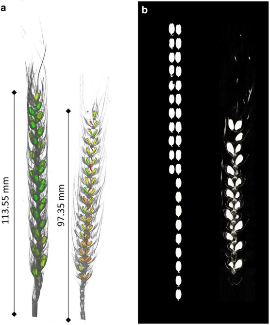
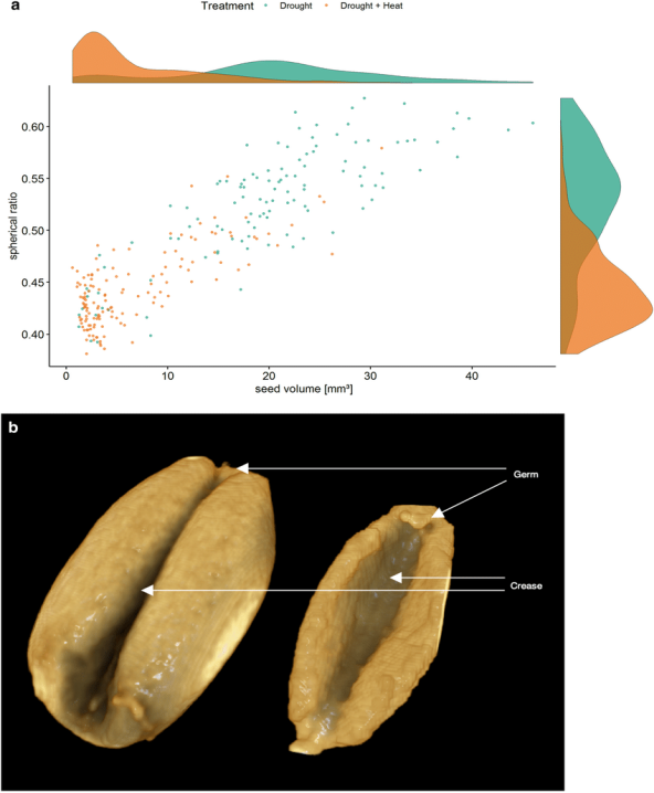
Background: Improving abiotic stress tolerance in wheat requires large scale screening of yield components such as seed weight, seed number and single seed weight, all of which is very laborious, and a detailed analysis of seed morphology is time-consuming and visually often impossible. Computed tomography offers the opportunity for much faster and more accurate assessment of yield components. Results: An X-ray computed tomographic analysis was carried out on 203 very diverse wheat accessions which have been exposed to either drought or combined drought and heat stress. Results demonstrated that our computed tomography pipeline was capable of evaluating grain set with an accuracy of 95-99%. Most accessions exposed to combined drought and heat stress developed smaller, shrivelled seeds with an increased seed surface. As expected, seed weight and seed number per ear as well as single seed size were significantly reduced under combined drought and heat compared to drought alone. Seed weight along the ear was significantly reduced at the top and bottom of the wheat spike. Conclusions: We were able to establish a pipeline with a higher throughput with scanning times of 7 min per ear and accuracy than previous pipelines predicting a set of agronomical important seed traits and to visualize even more complex traits such as seed deformations. The pipeline presented here could be scaled up to use for high throughput, high resolution phenotyping of tens of thousands of heads, greatly accelerating breeding efforts to improve abiotic stress tolerance.
Semiautomated 3D Root Segmentation and Evaluation Based on X-Ray CT Imagery
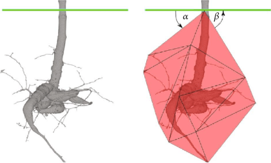
Background: Computed X-ray tomography (CTX) is a high-end nondestructive approach for the visual assessment of root architecture in soil. Nevertheless, in order to evaluate high-resolution CTX data of root architectures, manual segmentation of the depicted root systems from large-scale volume data is currently necessary, which is both time consuming and error prone. The duration of such a segmentation is of importance, especially for time-resolved growth analysis, where several instances of a plant need to be segmented and evaluated. Specifically, in our application, the contrast between soil and root data varies due to different growth stages and watering situations at the time of scanning. Additionally, the root system itself is expanding in length and in the diameter of individual roots. Objective: For semiautomated and robust root system segmentation from CTX data, we propose the RootForce approach, which is an extension of Frangi's "multi-scale vesselness" method and integrates a 3D local variance. It allows a precise delineation of roots with diameters down to several μm in pots with varying diameters. Additionally, RootForce is not limited to the segmentation of small below-ground organs, but is also able to handle storage roots with a diameter larger than 40 voxels. Results: Using CTX volume data of full-grown bean plants as well as time-resolved (3D + time) growth studies of cassava plants, RootForce produces similar (and much faster) results compared to manual segmentation of the regarded root architectures. Furthermore, RootForce enables the user to obtain traits not possible to be calculated before, such as total root volume (Vroot), total root length (Lroot), root volume over depth, root growth angles (θmin, θmean, and θmax), root surrounding soil density Dsoil, or form fraction F. Discussion. The proposed RootForce tool can provide a higher efficiency for the semiautomatic high-throughput assessment of the root architectures of different types of plants from large-scale CTX. Furthermore, for all datasets within a growth experiment, only a single set of parameters is needed. Thus, the proposed tool can be used for a wide range of growth experiments in the field of plant phenotyping.
Exploring Flood Filling Networks for Instance Segmentation of XXL-Volumetric and Bulk Material CT Data
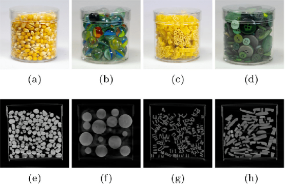
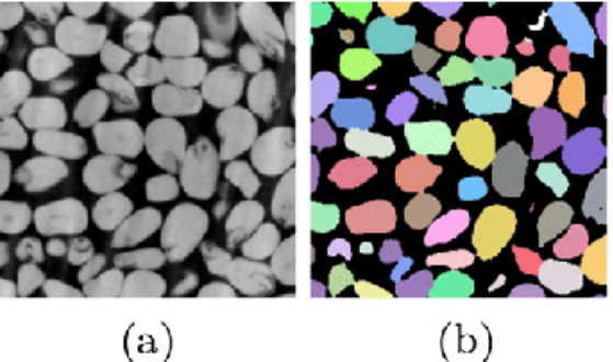
XXL-Computed Tomography (XXL-CT) is able to produce large scale volume datasets of scanned objects such as crash tested cars, sea and aircraft containers or cultural heritage objects. The acquired image data consists of volumes of up to and above $$\hbox {10,000}^{3}$$ 10,000 3 voxels which can relate up to many terabytes in file size and can contain multiple 10,000 of different entities of depicted objects. In order to extract specific information about these entities from the scanned objects in such vast datasets, segmentation or delineation of these parts is necessary. Due to unknown and varying properties (shapes, densities, materials, compositions) of these objects, as well as interfering acquisition artefacts, classical (automatic) segmentation is usually not feasible. Contrarily, a complete manual delineation is error-prone and time-consuming, and can only be performed by trained and experienced personnel. Hence, an interactive and partial segmentation of so-called “chunks” into tightly coupled assemblies or sub-assemblies may help the assessment, exploration and understanding of such large scale volume data. In order to assist users with such an (possibly interactive) instance segmentation for the data exploration process, we propose to utilize delineation algorithms with an approach derived from flood filling networks. We present primary results of a flood filling network implementation adapted to non-destructive testing applications based on large scale CT from various test objects, as well as real data of an airplane and describe the adaptions to this domain. Furthermore, we address and discuss segmentation challenges due to acquisition artefacts such as scattered radiation or beam hardening resulting in reduced data quality, which can severely impair the interactive segmentation results.
X-Ray CT Phenotyping Reveals Bi-Phasic Growth Phases of Potato Tubers Exposed to Combined Abiotic Stress
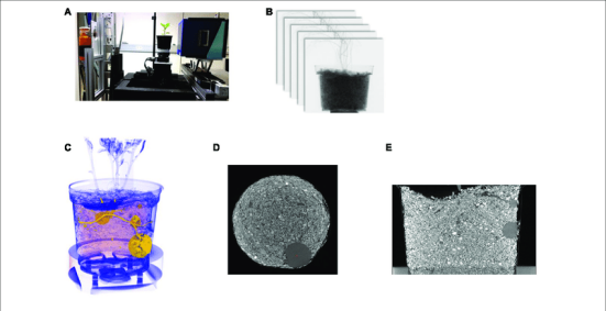
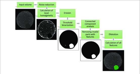
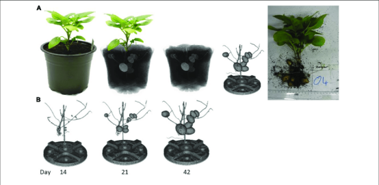
As a consequence of climate change, heat waves in combination with extended drought periods will be an increasing threat to crop yield. Therefore, breeding stress tolerant crop plants is an urgent need. Breeding for stress tolerance has benefited from large scale phenotyping, enabling non-invasive, continuous monitoring of plant growth. In case of potato, this is compromised by the fact that tubers grow belowground, making phenotyping of tuber development a challenging task. To determine the growth dynamics of tubers before, during and after stress treatment is nearly impossible with traditional destructive harvesting approaches. In contrast, X-ray Computed Tomography (CT) offers the opportunity to access belowground growth processes. In this study, potato tuber development from initiation until harvest was monitored by CT analysis for five different genotypes under stress conditions. Tuber growth was monitored three times per week via CT analysis. Stress treatment was started when all plants exhibited detectable tubers. Combined heat and drought stress was applied by increasing growth temperature for 2 weeks and simultaneously decreasing daily water supply. CT analysis revealed that tuber growth is inhibited under stress within a week and can resume after the stress has been terminated. After cessation of stress, tubers started growing again and were only slightly and insignificantly smaller than control tubers at the end of the experimental period. These growth characteristics were accompanied by corresponding changes in gene expression and activity of enzymes relevant for starch metabolism which is the driving force for tuber growth. Gene expression and activity of Sucrose Synthase (SuSy) reaffirmed the detrimental impact of the stress on starch biosynthesis. Perception of the stress treatment by the tubers was confirmed by gene expression analysis of potential stress marker genes whose applicability for potato tubers is further discussed. We established a semi-automatic imaging pipeline to analyze potato tuber delevopment in a medium thoughput (5 min per pot). The imaging pipeline presented here can be scaled up to be used in high-throughput phenotyping systems. However, the combination with automated data processing is the key to generate objective data accelerating breeding efforts to improve abiotic stress tolerance of potato genotypes.
