品质至上,客户至上,您的满意就是我们的目标
当前位置: 首页 > 新闻动态
科学家利用Videometer多光谱成像系统发表叶绿素荧光图像定量研究文章
发表时间: 点击:802
来源:北京博普特科技有限公司
分享:
最近,科学家利用VideometerLab多光谱成像系统,发表了题为“Quantification of chlorophyll fluorescence in soybean seeds by multispectral images and their relationship with physiological potential”,文章发表于期刊J. Seed Sci. 44•2022。
大豆种子叶绿素荧光的多光谱图像定量及其与生理电位的关系
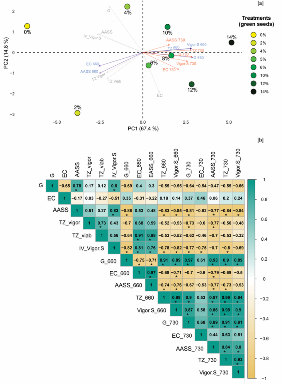
研究了检测大豆种子叶绿素荧光(CF)的多光谱图像分析技术,以评估CF信号与种子生理潜力之间的关系。用8种处理,分别对应0%、2%、4%、6%、8%、10%、12%和14%的绿色种子,对BMX Desafio RR 8473 RSF和96R10 IPRO两个通过不同种子质量测试的品种进行处理。最初,使用660 nm和730 nm光谱测定种子的CF,然后对同一种子进行发芽、电导率、饱和NaCl溶液加速老化、四氮唑和计算机化幼苗图像分析(活力-S)测试。采用完全随机设计,以及每个处理重复。使用R®软件对发芽、活力和CF测试的数据进行方差分析(ANOVA),并通过Scott Knott测试对均值进行分组(p≤ 0.05). 计算评估中所有组合的皮尔逊线性相关系数(r),通过t检验确定r值的显著性(p≤ 0.05),并对主成分进行多变量分析。绿色种子比例的增加有助于叶绿素荧光信号的增加,并与种子生理质量负相关;样品中绿色种子含量超过4%会导致生理潜力显著下降。因此,通过多光谱图像检测到的叶绿素荧光与大豆种子的生理潜力呈负相关。
索引项:
叶绿素保留;甘氨酸(L.)Merrill;绿色种子;种子活力;光谱学

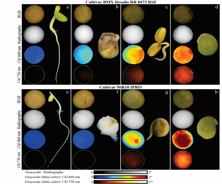
Figure 3 Soybean seeds with increasing levels of retained chlorophyll, obtained from RGB (Red-Green-Blue) images of internal morphology with the same seeds observed in radiography and chlorophyll fluorescence signals detected by multispectral images at 660 nm (Chl 660 nm) and 730 nm (Chl 730 nm), respectively. In the multispectral images of the soybean seeds, transformed into color map based on gray levels, the hues of the colors dark blue (660 nm, of VideometerLab) and black (730 nm, of SeedReporter) represent the seeds with “yellow” seed coat and embryo, whose chlorophyll was degraded and, for that reason, have low chlorophyll fluorescence signals; the hues with a tendency toward intense red indicate the “greenish” seeds, whose chlorophyll is retained in the seed coat and embryo and, therefore, express greater signals of chlorophyll fluorescence. a; e: normal soybean seedling resulting from yellow seed; b; f: dead seed, with mechanical damage, covered by fungal mycelium and weak signals of chlorophyll fluorescence; c; g: abnormal soybean seedling originating from seed with partial chlorophyll retention; d; h: dead seed resulting from green coloring, which indicates a high level of retained chlorophyll.
Quantification of chlorophyll fluorescence in soybean seeds by multispectral images and their relationship with physiological potential
Abstract
The multispectral image analysis technique to detect chlorophyll fluorescence (CF) in soybean seeds was studied to assess the relationship between CF signals and seed physiological potential. Eight treatments, corresponding to 0%, 2%, 4%, 6%, 8%, 10%, 12%, and 14% green seeds, were used on two cultivars, BMX Desafio RR 8473 RSF and 96R10 IPRO, which passed through different seed quality tests. Initially, the CF of the seeds was determined using 660 nm and 730 nm spectra, and then the germination, electrical conductivity, accelerated aging with saturated NaCl solution, tetrazolium, and computerized seedling image analysis (Vigor-S) tests were performed on the same seeds. A completely randomized design was used, as well as replications of each treatment. Analysis of variance (ANOVA) was performed on the data from germination, vigor, and CF tests using the R® software, and the means were grouped by the Scott-Knott test (p ≤ 0.05). Pearson’s linear correlation coefficients (r) were calculated for all combinations among the evaluations with significance of the r values determined by the t-test (p ≤ 0.05), and multivariate analysis of the principal components was performed. Proportional increases in green seeds contribute to an increase in chlorophyll fluorescence signals and have a negative correlation with seed physiological quality; levels above 4% green seeds in the samples result in marked losses in physiological potential. Therefore, the chlorophyll fluorescence detected through multispectral images is inversely related to the physiological potential of soybean seeds.
Index terms:
chlorophyll retention; Glycine max (L.) Merrill; green seeds; seed vigor; spectroscopy
